
The Pathologist’s challenge is to identify the disease based on sometimes vague morphological features of the tissue. During last decade, digital pathology entering the field, rapidly changing the clinical routines. Whole slide images (WSI) became a standard for the diagnostic digital pathology nowadays.
With modern technology though comes new challenges: how to to store large amounts of data generated by slide scanners, what to do with physical slides after digitalisation, how to ensure access to the data for all the pathologists but keep the data protected and safe?
Characteristics of Whole-Slide Images
Image acquisition
Most optical microscopes have an eyepiece which provides 10X magnification and maximal objective lens with 40X magnification which results in 400X scale view – making it possible to distinguish features of the side of .25 microns — that’s about the thickness of one human hair. Digital image of the tissue sample can be generated by microscopes equipped with optical cameras and even by smartphones. However, good-quality hight resolution WSI acquisition requires specifically designed instrument – slide scanner.
Image dimensions and data size
Clinical tissue samples can be up to 50mm x 25 mm in size, and for the diagnostic needs they need to be digitised with a resolution of .25 microns/ pixel. However, sometimes even larger samples are used and they should be digitalised with the same, high resolution.


Image visualisation
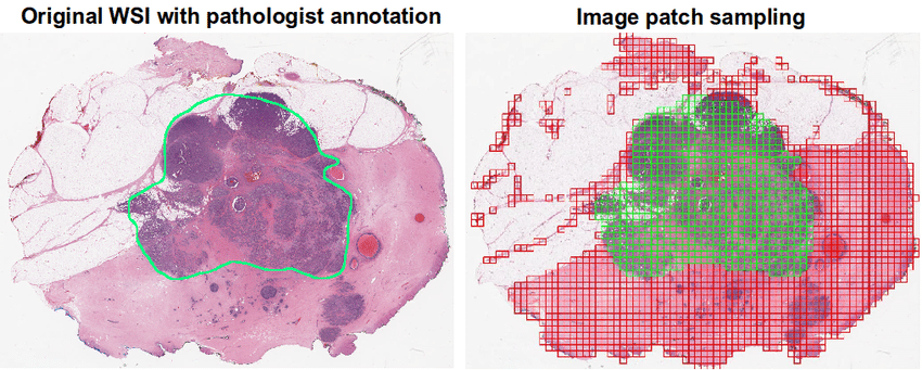
To make the advantage of novel technologies, pathologists need to be able to access the WSIs, rapidly pan and zoom images, move from regions to region, and observe different zones of the diagnostic sample. There is a number of venders providing WSI viewing, and often also file management systems. These viewing systems often offer the possibility to label or annotate the WSIs, allowing data sharing between pathologists, which may be needed for example, if difficult clinical cases, that require consensus of several doctors.
Image storage infrastructure
The DICOM standard is a way to exchange medical images, and it has been widely used in hospitals around the world. This format offers both storage and forward architecture as well interactive access between a client workstation and a server. The image data is stored in a tiled fashion to facilitate rapid panning. This enables random access and synthesis at any desired resolution without loading large amounts of unnecessary data, which can be saved on disk for later use when zooming or focusing closer than what’s currently visible.
To help with zooming, an image can be stored at several pre-computed resolutions. This enables synthesis of subregions and their variations without scaling large amounts of data!