

Artificial Intelligence (AI) has drastically changed the medical field in recent years. One of the most useful applications of this technology is medical image annotation. With its help, Histoone has started a new project for cancer detection. Here is a case study of how Histone started using AI annotation in lung cancer detection.
Introduction to the project
We had a need to develop a model for lung cancer detection. This is where an app can be stepped in. Diagnosis of lung cancer is a very difficult process, especially when it’s in advanced stages.
The need to develop a model for lung cancer detection. For better treatment and prognosis, we need biopsy analysis with automatic AI annotation.
Early detection and diagnosis of cancer are critical for increasing the chances of successful treatment. However, the process of identifying and diagnosing cancer is often a lengthy and complicated one. Physicians must carefully examine biopsy samples under a microscope in order to make a diagnosis. This process is time-consuming and can be subject to human error.
In order to speed up the process of identifying and diagnosing lung cancer, we have used a new app that uses artificial intelligence (AI) and machine learning techniques to analyze and annotate biopsy images.
The process of AI medical image annotation
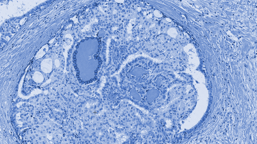
- The process starts by loading in images and labeling them for training.
- The dataset for the training session of the model needs to be created. In order to create a dataset for the training session of an AI medical image annotation model, the slides need to be labeled accordingly.
- AI labels the image slides for lung cancer detection.
- data with annotations must be exported in a format that is compatible with the machine learning software being used.
- Finally, the model must be trained on the dataset in order to predict the correct labels for new images.
Once the model has been trained, it can be used to detect lung cancer with high accuracy on new patient slides without human input!
Cloud-based system
The process begins with a team of pathologists working on the project together. However, due to geographical constraints, they are unable to physically collaborate in one place. Therefore, they rely on cloud-based data stores that provide secure access and protection against data loss or theft from outside sources.
Working in cloud services allowed us not only greater accessibility but also more efficiency since we’re able to work with our team members all over the world.
The cloud-based datastore also ensures that all information is safe and secure.
Iteration of the labeling-training rounds.
The next step is the iterative process of labeling-training rounds. After each round of training models is validated, the annotations can be improved based on the results before moving onto the next round. This process helps ensure accuracy by allowing teams to identify any areas where additional information needs to be added or adjusted accordingly.
Today, cancer is one of the leading causes of death worldwide. This app is designed to streamline the process of digital pathology, making it more efficient and accurate. With this app, our physicians were able to quickly and easily identify cancerous cells, leading to improved patient outcomes.
Why medical data annotation is important for businesses?
AI medical image annotation can save businesses time and money by eliminating the need for manual labor associated with labeling images. Additionally, it can also help reduce errors because it relies on computer algorithms rather than human input alone. This means that companies can trust that their data is accurate and up-to-date without having to double-check every entry manually. By using AI medical image annotation, businesses can streamline their operations while also ensuring accuracy and precision in their workflows.
AI medical image annotation provides businesses with an efficient way to label images quickly and accurately without requiring manual input from employees or other external sources. Not only does this save time and money but also ensures accuracy in data collection processes while protecting against potential data loss or theft from outside sources.
Please have a look at the demo